We have the know-how tools and techniques along with the expertise to bring 3D medical conditions to life on screen through 3D animation, so patients and care providers like Physicians & surgeons, medical students & professionals around the world can better understand the presented topic and material. With the blend of different mediums such as 3D animation, virtual reality, voiceover, and live-action video, we can narrate a story that resonates with your target audience. We serve the purpose of raising awareness for your cause using our 3D Heart animation services.
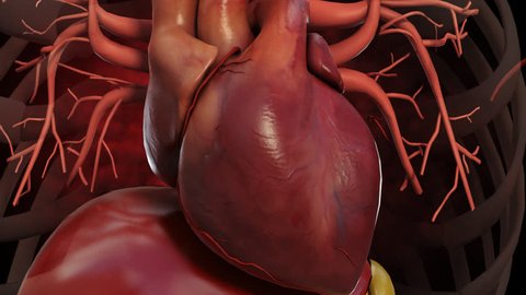
This 3D heart animation video does a great job of representing the cycles through the human heartbeat.
It is interesting to note that subtle twist the heart in 3D animation makes when it beats as well as the enlargement of the arteries as the blood pushes through. The human heartbeat in 3D animation mostly does a great job of displaying the basic human heartbeat through an interactive heart.
This heart animation is created from a 3D heart model which is very difficult to create a very precise 3d model of the heart & how the heart works because the heart never stops moving when you take a CAT scan or an MRI. It took an enormous amount of time and effort in developing the 3d heart model, building it according to the anatomy of the heart & the best data available on heart flow, heart physiology, the blood supply of heart & circulation of blood through the heart, heart pumping blood, cardiac blood flow, cardiac circulation, how the heart works, etc. in the form of videos, motion images, animations, textures, etc.
What is Heart in 3D animation? Or what does the Heart look like and how does the heart works?
The heart is mainly composed of specialized cardiac muscle, and it is four-chambered, with a right atrium and ventricle, and an anatomically separate left atrium and ventricle. The blood flows from the systemic veins into the right atrium, subsequently to the right ventricle, from which it is pumped to the lungs, then returned into the left atrium, then to the left ventricle, from which it is driven into the systemic arteries.
It will be easier to say, the heart is therefore functionally composed of two hearts: one is the right heart and the other is the left heart. The right heart consists of the right atrium, which receives deoxygenated blood from the body, and the right ventricle which pumps it to the lungs under low pressure; and the left heart, comprising of the left atrium, which receives oxygenated blood from the lung, and the left ventricle, which pumps it out. The muscle that pumps blood through the heart shown in animation received from veins into arteries throughout the body. It is located in the chest behind called the sternum (breastbone; in front of the trachea, esophagus, and aorta; and above the diaphragm muscle that separates the chest and abdominal cavities). The usual or the normal heart is about the size of a closed fist and weighs about 10.5 ounces. The shape of the heart is a cone-shaped, with the point of the cone pointing down to the left. Two-thirds of the heart lies in the left side of the chest with the balance in the right chest.
The heart starts beating 22 days after conception and continuously pumps oxygenated red blood cells and nutrient-rich blood and other compounds like platelets throughout your body to sustain the life of your organs. The pumping power of the heart also pushes blood through organs like the lungs to remove waste products like CO2 from the body. The heart called a fist-sized powerhouse that beats (expands and contracts) about 100,000 times per day, pumping five or six quarts of blood each minute, or about 2,000 gallons per day.
In general, if the heart stops beating, in about 4-6 minutes of no blood flow circulation, brain cells begin to die and after 10 minutes of no blood flow circulation, the brain cells will certainly cease to function and effectively be dead. There are little exceptions to the above.
In 3D Heart animation, how the heart works are demonstrated through a regulated series of events that cause this muscular organ to contract (squeeze to push blood) and then relax (refill with blood).
Usually, the normal heart is said to have 4 chambers that undergo the squeeze and relax cycle at the specific course of time intervals that are regulated by a normal sequence of electrical signals that arise from specialized tissue.
In addition to this, the normal sequence of electrical signals can be sped up or slowed down depending on the needs of the individual, for example, the heart will automatically rise in speed its electrical signals to respond to a person running and will automatically slow down when a person takes a nap or a while sleep.
This 3D Heart animation, 3D heart anatomy animation, human blood circulation system animation, heart beating video animation, heart pumping blood, heart function animation, heart flow video is designed to help individuals learn the heart anatomy and circulatory system, and provide some vision about heart health. It is not designed to present the many problems that can occur with the heart to the body under high pressure.
Heart Flow 3D Animation explains how does blood flow through the right and left side of the heart?
3D Heart Animation of the blood flow through the right and left sides of the heart work together is easy to understand. The pattern described below is repeated over and over again (heart rhythm or heartbeat), causing blood to flow continuously to the heart, lungs, and body to supply oxygen and nutrients to the human body cells and to deliver waste products to organs that eliminate them from your body. It is as universal, veins return with blood carrying CO2 whereas arteries usually contain O2 enriched red blood cells. However, the blood flow through the heart is a little different, is manifested in this blood flow 3D animation. For example:
The right side of the heart animation
Blood makes entry into the heart through two large veins,i.e. one is inferior and the other is superior vena cava, which removes out oxygen-poor blood from the body into the right atrium of the heart.
As the atrium contracts, blood supply flows from your right atrium into your right ventricle through the open tricuspid valve.
When the ventricle is full, the tricuspid valve shuts completely. Thus, at the time of the contraction of the ventricle, the blood is prevented from flowing backward into the atria.
As the ventricle contracts, blood leaves or flows from the heart through the pulmonic valve, into the pulmonary artery and to the lungs where it gets oxygenated. Noticing the fact that oxygen-poor or CO2 containing blood goes through the pulmonary artery to the lungs where CO2 is exchanged for O2.
The Left side of the heart (operating simultaneously as the right side of the heart) animation
The pulmonary vein empties oxygen-rich blood from the lungs& pumps into the left atrium of the heart.
As the atrium contracts, blood supply flows from your left atrium into your left ventricle through the open mitral valve.
When the ventricle is full, the mitral valve shuts completely. Thus, at the time of the contraction of the ventricle, the blood is prevented from flowing backward into the atrium.
As the ventricle contracts, oxygen-enriched blood leaves the heart through the aortic valve, into the aorta, and to the arteries, and eventually into veins to complete the blood circulation system in your body.
Normal Heart anatomy and Heart physiology 3D Animation
3D Animation effectively explains the concept of Normal heart anatomy and physiology that needs the atria and ventricles to work chronologically, contracting and relaxing to blood pumping out of the heart and then to let the chambers refill. When blood leaves each chamber of the heart, it passes through a valve that is designed to prevent backward flow or circulation of blood.
There are four heart valves inside the heart:
- Mitral valve in the middle of the left atrium and left ventricle
- Tricuspid valve in the middle of the right atrium and right ventricle
- Aortic valve between the left ventricle and aorta
- Pulmonic valve (also called pulmonary valve) between the right ventricle and pulmonary artery
How do the heart valves work in 3D animation?
3D animation of the heart valves work shown here the same way as one-way valves in the plumbing of your home works. They prevent blood from flowing (circulation of blood) in the incorrect direction.
- Each valve contains a set of flaps, called leaflets or cusps.
- The mitral valve has two leaflets; the others have three leaflets.
- The leaflets are adjoined to and supported by a ring of tough, fibrous tissue called the annulus.
- The annulus helps to maintain the appropriate shape of the valve.
- The leaflets of the mitral and tricuspid valves are also sustained by tough, fibrous strings termedas chordae tendineae.
These are alike to the strings supporting a parachute. They enlarge from the valve leaflets to small muscles, called papillary muscles, which are a portion of the inside walls of the ventricles.
Role of 3D Medical Heart Animation
Heart Beats: Through 3D medical Heart animation every minute movement of the heart in action is visible by setting the heart rate to a simulation of real scenarios or could be controlled by the 3D animation.
Systems:3D medical Heart animation explores body systems linked to the heart for a greater synopsis of how the body functions.
Path of Origin: To find an artery back to its origin at the heart then explore or examine the other structures within the path by isolation or highlight of the path.
Thorough view: Digging into the heart anatomy through3D animation or video magnifies the blood vessels model to find out the fine layers and features of the vessels like never seen before.
Animations: Virtually visualize pathologies and procedures with comprehensive3D medical video animations. 3D Heart animations help to realize how conditions develop and are treated.
Screens: Heart in 3D animation helps the audience to sight the clinical or medical screens, featuring the customized model setups for analyzing conditions as well as the contents of complementary texts, visual graphics & imagery.
Courses: Complete breakdown analysis of very complex cardiac topics by 3D models of Heart animation, cardiac circulation animation, cardiac blood flow, etc. animation becomes a lesson of guidance to medical experts and other audiences.
Introduction to Anatomy of the Heart through 3D Animation
The anatomy of the heart in 3D animation is designed to give you an all-inclusive introduction of the heart. The interactive heart and the 3D model covering the concepts and principles related to the human gross anatomy of the heart covers it all.
ANATOMY– The topic of the heart can be chosen quickly and easily with screens and recordings according to the anatomical structure.
PHYSIOLOGY– Heart Physiology animation explains how the heart functions concerning the rest of the body along with extra detail of highlighting text and images.
ANNOTATE– Adding labels to mark the diagram or drawing on the model or structure of the custom diagrams for future reference can be one of the unique qualities in 3D medical heart animation.
EDIT – Using Tools you can change the heart structure or model to illustrate the conditions or dig in deeper areas of the organ to explain in detail.
SAVE– You can make a recording of your custom model set up of heart in 3D animation to explain the key points of it.
ACCURATE– The product or the model of Heart in 3D animation has to look stunning, medically accurate, clean, and organized along with comprehensive to understand.
Why we have been in the industry standard for so many years?
Using our 3D anatomy archive, you will be able to explore & discover any anatomical model you need to illustrate or animate, and within minutes, you’ll be able to begin creating beautiful, photo-realistic renderings of the heart animation.
The blend of medical precision and high aesthetic quality makes heart anatomy, heart animation, heart physiology, cardiac blood flow animation, human blood circulation animation & other concepts of the heart to be artistic & accurate through 3D.
With our clean geometry and organized hierarchy, you will be able to quickly drill down to the focus of your production and create amazing rendering visuals illustrating exactly what you want to be explained by us through the model.
This is the world’s most popular interactive virtual anatomy of the heart in 3D animation is made available to you as a platform on which you can build.
The market is volatile & competitive but at the same time, the product like this is easily adaptable by the customers due to advanced medical technology& fully 3D interactive power to captivate the viewers’ attention.
We all are living in 3D medical animation and interactive Augmented Reality, Mixed Reality and Virtual Reality applications with an actual emphasis on the pharmaceutical, medical, and biotechnological sectors have been very vital for us. Our main goal is to make medical education fascinating, discoverable, and creative fun for physicians, students as well as accessible to patients – anywhere and anytime, on places, medical offices, hospitals, available to the classroom, lecture hall or living room etc. We are avid about taking medical education one step ahead and have developed a visually stunning and highly interactive content based on medical and scientific specifications as in real life.
Using 3D cardiology animation in a medical video helps explain complex vascular structures, and demonstrates the interdependent functions of the four heart valves mentioned above: the aortic, mitral, pulmonary, and tricuspid. Watching a 3D heart animation is easier for the brain to pick faster because the way that all of the parts of the heart work together is demonstrated in these cardiac animations. A heart 3D animation describes the interdependencies of the heart and the circulatory system in a more dynamic way than viewing only a still heart medical illustration or an image.
The advantage of creating a circulatory system 3D animation is that it can capture any single frame as a high-resolution image. Heart physiology or heart anatomy animation runs at 30 frames per second technically. This calculates that for each minute of cardiovascular animation there are 1,800 static images to choose from to create medical diagrams of the heart. Many clients use both the 3D animation of the heart medical illustrations, cardiovascular system animations, helps explain the patients, physicians & other medical experts & students about the concepts of what insistent implants, balloon catheters, heart-valve replacements, automatic external defibrillators, pharmaceutical products for cardiology treatments, and robotic-assisted surgery in their presentations or display them on internal website case study in an interactive manner. Adapting to medical 3D animation medical students & patients can comprehend in before to how the surgery works, what the benefits are, and how the surgeon performs the procedure.
From a decade or so, our team works on our client terms, vision& ideas that translate their need into a rich 3D experience of virtual cardiology animation or 3D Heart animation. We will collaborate with you to support your medical device marketing, build integrated media solutions, and supply you with the creative assets based solutions needed for your next future campaign in the medical world of technology.
Are you looking for someone to create a 3D 4K Heart Animation Video for your business?
Contact us or request a demo with us. We would be more than happy to help you.
